Futurum offers a unified, high-fidelity model that supports entire procedural training—from diagnostics to surgery—with anatomical realism and soft tissue fidelity all in one model.
1.
Replaces cadavers with a realistic, ethical alternative
2.
Provides comprehensive procedural training
3.
Enhances anatomical and diagnostic understanding
4.
Delivers realistic sensory feedback
5.
Skill mastery through repetition and consistency
6.
Supports future-ready learning
7.
Increases accessibility and standardisation
8.
Enables structured, step-by-step skill progression
9.
Reduces training costs and resource dependency
Veterinary dentistry is facing a critical skills gap on a global scale. Out of more than 600,000 veterinarians worldwide, fewer than 350 are board-certified dental specialists across both the American and European Veterinary Dental Colleges.
There are 900 million pet dogs and 370 million pet cats worldwide. Periodontal disease occurs in 85% of dogs over the age of 2 and 75% of cats over the age of 3, making it the most common disease of both dogs and cats. With a global shortage of veterinary dentists, non-cadaveric models offer a flexible solution to improve access to training.
Access to cadaveric specimens for training is limited, expensive, and often ethically and logistically challenging. Our anatomically accurate, 3D-printed models solve this by enabling safe, repeatable, and risk-free skills development—even for rare and complex pathologies.
With applications that will extend across all areas of canine and feline anatomy, and potential to scale into other species, our technology offers a revolutionary path forward in veterinary education and advanced clinical training.
Historically, training has relied on cadavers—a resource that is costly, increasingly difficult to access, and presents hygiene and biosecurity risks. Growing concerns around ethical practices and sustainability in a time where we have the tools to produce a modern alternative that delivers the same realism and educational value.
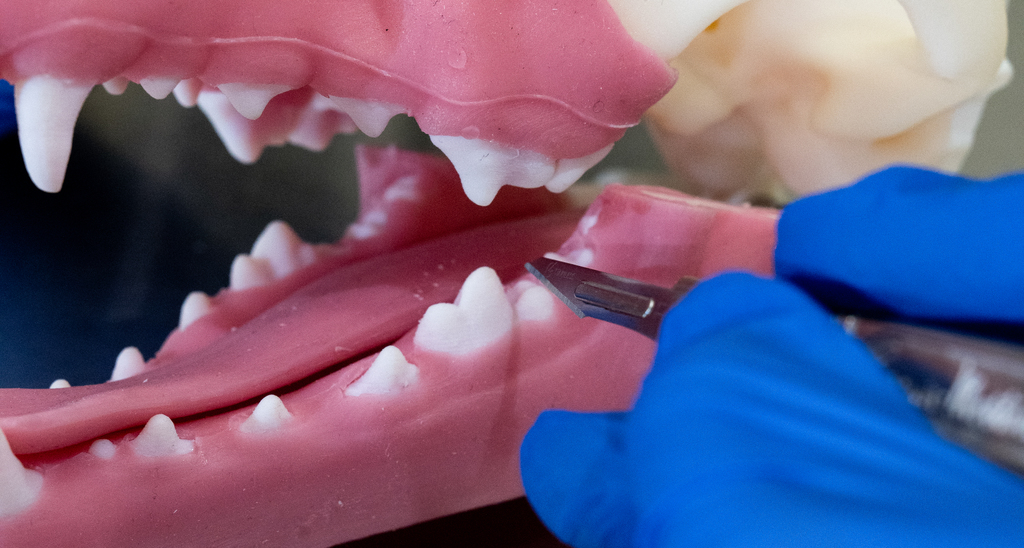
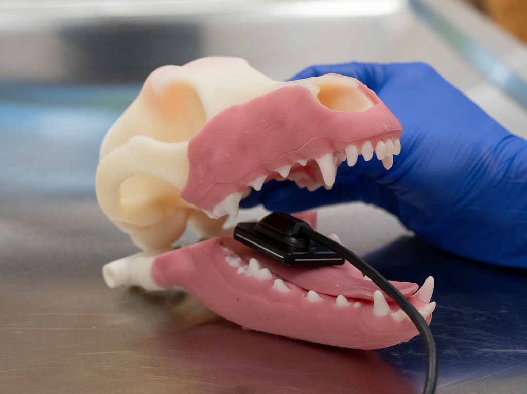
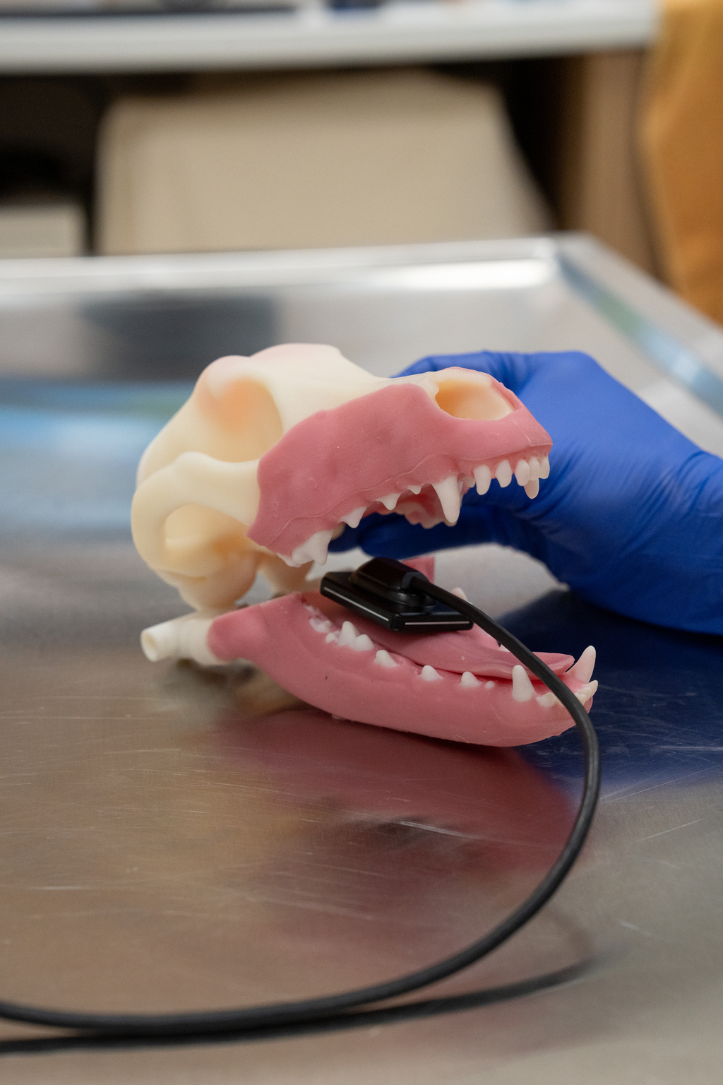
Futurum Animal Biotech has developed a realistic, reusable, and designed to replace cadavers while enhancing the quality and consistency of veterinary training. By replicating the look, feel, and function of real canine oral anatomy, the model provides a standardised, accessible, and ethically sound solution that supports skill development for students and professionals.
Every detail of the Futurum model was developed with precision—from digital design to physical prototype. The anatomy was mastered at an internal level, ensuring that structures beneath the surface are just as accurate as what’s seen externally.
Using advanced anatomical data, engineering expertise, and clinical insight, we transformed a virtual concept into a high-fidelity, lifelike training tool.
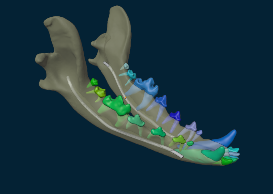
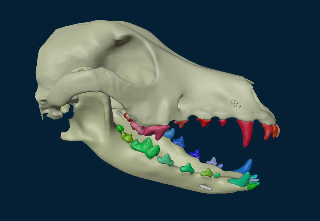
Engineered to integrate a complete endodontic system and to accurately replicate the movement and function of the gingival attachment. This allows for the creation of realistic mucoperiosteal flaps and key anatomical structures such as the periodontal ligament, mandibular canals, and nerve foraminee.
Analysis of the Futurum non-cadaveric model anatomy:
of participants responded that they would be interested in using non-cadaveric models in their own clinical practice for training and skill development.
of participants rated the model touch and feel 5/5
of participants rated the model touch and feel 4/5
of participants responded that they believe the use of the non-cadaveric model is important to improving ethics and sustainability within veterinary training.
While other non-cadaveric market offers impressive modular tools for focused skills (like radiographs or endodontics), it is common to require an entirely separate model for each application.
Futurum offers a unified, high-fidelity model that supports entire procedural training—from diagnostics to surgery—with anatomical realism and soft tissue fidelity all in one model.
Designed with precise and complete anatomical fidelity, including soft tissue, jaw structure, tooth anatomy and full dentition replicating the entire oral cavity for comprehensive training.
Primarily skeletal or dental-focused with underdeveloped or no soft tissue representation; often lack realistic anatomical detail beyond functional structures, which limits the scope of veterinary training.
Differentiator: Futurum delivers complete anatomical accuracy focused on the oral cavity, innovated with detailed internal structures that replicate the cadaver experience- without the need for any ethical, biohazard or sourcing considerations.
Futurum’s anatomically complete, all-in-one model supports a full range of procedures in a single, unified platform. Built for durability and repeated use, it’s ideal for both foundational and advanced skill-building. By providing a complete intraoral environment, it allows for realistic, uninterrupted practice.
Other models on the market fragmented, relying on separate modules for different tasks. This piecemeal setup forces users to move between products to simulate what should be a continuous process. While helpful for isolated skills, these models lack the integration needed for complete procedure training, limiting their effectiveness in building clinical confidence and workflow continuity.
Differentiator: Delivers a true-to-life intraoral experience designed for full-procedure training, not just isolated steps, saving time and cost while improving training continuity. Built to support progressive learning from the ground up, it enhances operator outcomes by guiding users through a structured sequence: mastering radiology, understanding normal anatomy, performing nerve blocks, starting with simple extractions, then advancing to large gingival flaps and complex extractions. The model is intentionally designed without replaceable parts so the training experience has the correct flow, ensuring comprehensive skill development and ultimately improving success in patient procedures.
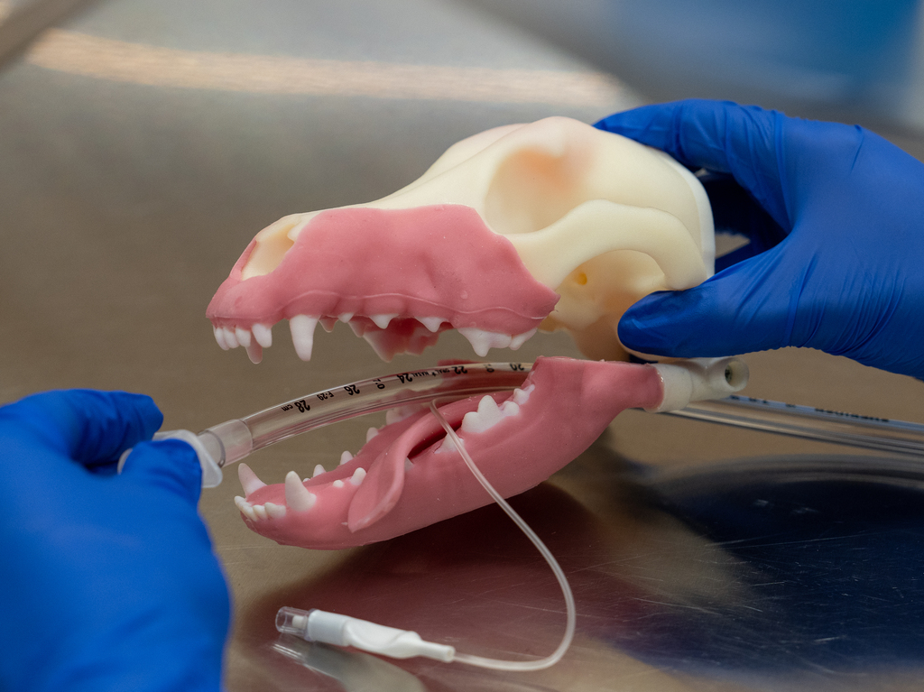
Designed to replicate a realistic experience of clinical procedures, the Futurum model delivers high-fidelity sensory feedback through its materials that mimic the texture, resistance, and responsiveness of actual soft tissue, bone, and dentition. The model allows users to experience realistic tactile cues during procedures like incisions, drilling, extractions and suturing, which are crucial for developing the skills required in veterinary dentistry.
Other models use uniform, low-fidelity materials that fail to reflect the variability and complexity of real oral anatomy. The absence of authentic tactile feedback limits the potential to develop these skills in training.
Differentiator: No model yet matches Futurum in its ability to deliver comprehensive procedure training while simultaneously replicating authentic sensory feedback and material realism.
Veterinary dentistry often lacks hands-on training due to limited cadaver access, hygiene and biosecurity concerns, and ethical issues. Traditional methods do not adequately prepare students for real clinical procedures. Futurum addresses this gap with a realistic and accessible alternative.
The model supports multiple essential procedures including:
Radiographic positioning
Regional anesthesia (nerve blocks)
Tooth extractions
Gingival surgery
Root canal therapy
Gingival flap creation and suturing
Extremely realistic. It replicates full oral anatomy including soft tissue (mucosa and periosteum), periodontal ligaments, mandibular canals, and vascular structures. It also offers tactile feedback similar to live tissue, critical for providing an accurate training experience.
The Futurum model is designed for multiple uses, allowing repeated training across a wide range of procedures without needing replaceable parts, making it cost-effective and sustainable.
Unlike fragmented modular systems, Futurum provides a complete intraoral environment in a single model. It allows full-procedure training—diagnosis to surgery—while delivering anatomical and tactile realism that other models cannot match.
Yes. It can generate radiographs that closely mimic true canine anatomy, showing detailed structures like the mandibular canal, inferior alveolar nerve, and pulp chambers—crucial for diagnostic learning.
Yes. The model is manufactured in Australia and can be customised for educational institutions or specialty training, with future iterations planned to simulate conditions like tumours, malocclusions, and deformities.
The model removes the need for cadaver sourcing, reducing ethical concerns and biohazard risks. It supports sustainable education by eliminating waste and the ethical complications of animal use in training.
Please fill out the 'register your interest' form, and we will be in contact to discuss the details of your order.